Continuing Education:
Endoscope Drying
This 1-hour program — available in person or via webinar — provides sterile processing professionals with essential knowledge on endoscope drying best practices.
What You'll Learn:
- ✓ WHY we need to dry endoscopes after reprocessing
- ✓ HOW to accomplish effective channel drying
- ✓ WHAT automation options exist to streamline the process
Expert Series
Monthly insights from industry experts on endoscope drying, infection prevention, and reprocessing best practices.
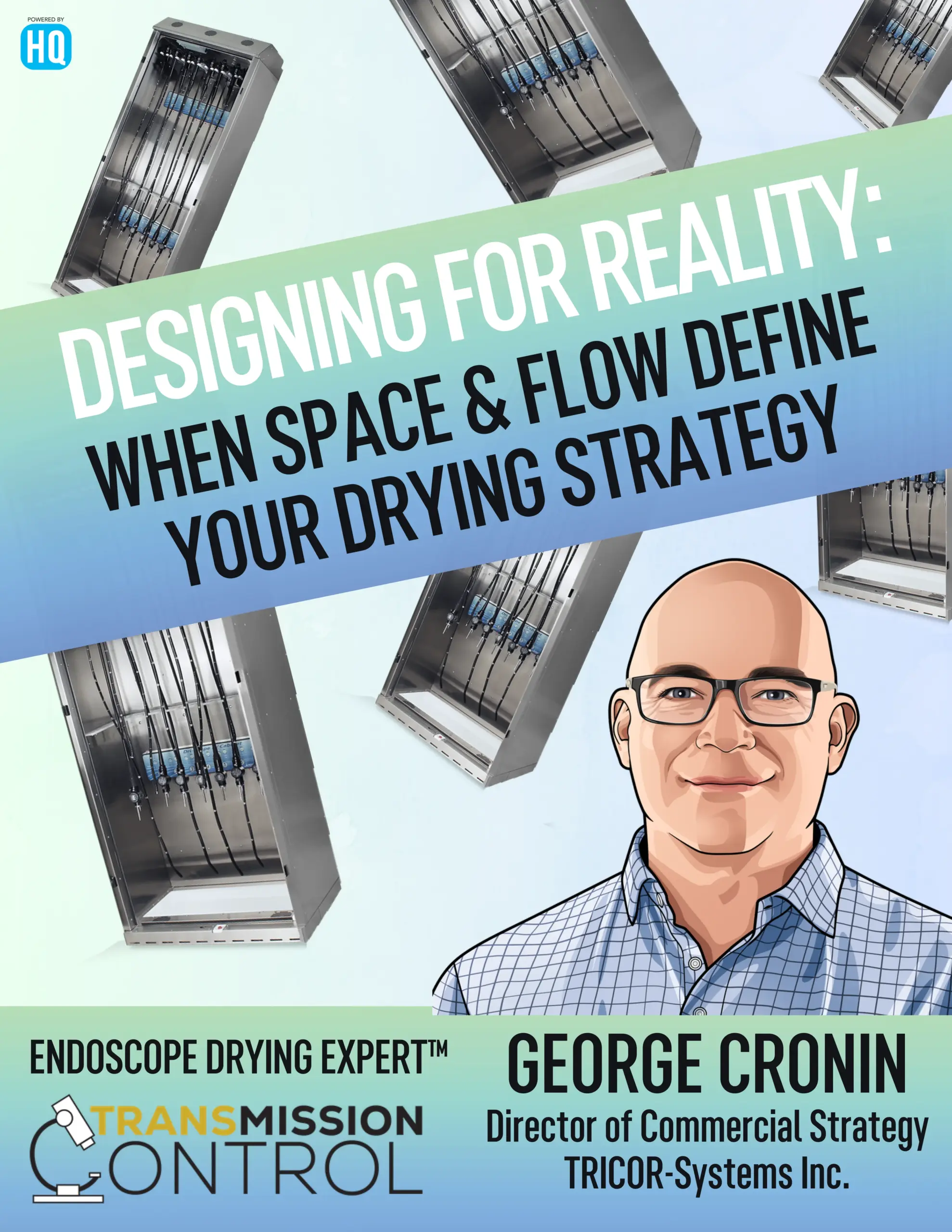
June 2026
This month's Expert Series explores where does drying happen in your department? Is the cabinet near the clean side, or down the hall? Do staff transport scopes across traffic patterns to complete the cycle? In high-volume environments, even short distances can create delays, increase handling, and introduce risk.
Featuring insights aligned with ANSI/AAMI ST91:2021 standards and real-world implementation strategies from leading healthcare facilities.
Read June 2026 Edition →Past Editions
Video Library
Product demonstrations, setup guides, and maintenance tutorials to help your team get the most out of Dri-Scope Aid® products.
Dri-Scope Aid® Jet~Stream

Quick Start Guide
Get up and running with your Jet~Stream in minutes
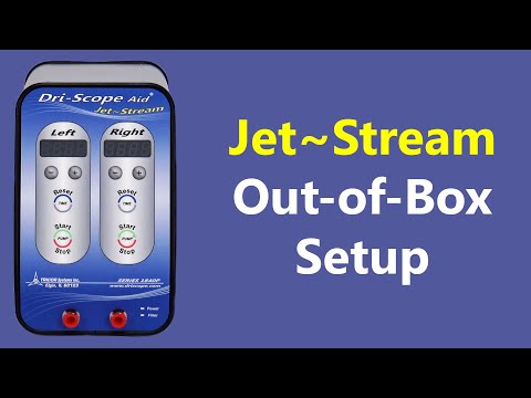
Out of the Box Setup
Complete unboxing and initial installation
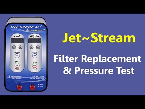
Pressure Test
How to perform routine pressure testing
Dri-Scope Aid® Tracker
Quick Start Guide
Setup Information
Pressure Test Demo
How to perform system pressure testing
Filter Replacement
Step-by-step filter replacement guide
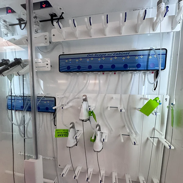
Dri-Scope Aid® Cabinet
Quick Start Guide
Features and operation walkthrough
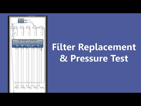
Filter Replacement Pressure Test
How to perform system pressure testing
Podcasts
Listen to industry discussions featuring Dri-Scope Aid® and the importance of endoscope drying.
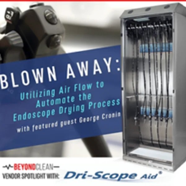
Beyond Clean: Vendor Spotlight
Join the Beyond Clean podcast for an in-depth conversation about the critical role of endoscope drying in infection prevention and how Dri-Scope Aid® is helping facilities meet compliance standards.